
CISN - How Cancer is Studied - Research Technology pg. 2
| You Are Here: Home > Cancer Research > How Cancer is Studied > Background > Research Technology pg. 2 |
||||||||||||
Research Technology - Page 2Fluorescence In Situ Hybridization (FISH)FISH is a technique used to detect and localize the presence or absence of specific DNA sequences on chromosomes. FISH uses fluorescent probes that bind to only those parts of the chromosome with which they show a high degree of sequence similarity. Fluorescence microscopy can be used to find out where the fluorescent probe is bound to the chromosome.
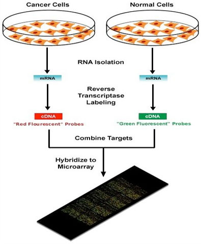
In medicine, FISH can be used to form or confirm a diagnosis, to evaluate a prognosis, or to evaluate remission of a disease, such as cancer. Treatment can then be specifically tailored to the specific situation. In reseach, FISH can be used to measure chromosome defects like gene deletions and duplications or fusions that can be associated with cancer. MicroarraysBecause this technique is so often discussed in research, this section contains a little more detailed information than the others. Background: With only a few exceptions, every cell of the body contains a full set of chromosomes and identical genes. Only a fraction of these genes are turned on or "expressed", however, and it is the subset that is "expressed" that confers unique properties to each cell type. "Gene expression" is the term used to describe the transcription of the information contained within the DNA, the repository of genetic information, into messenger RNA (mRNA) molecules; these molecules are then transformed into the proteins that perform most of the critical functions of cells. Scientists study the kinds and amounts of mRNA produced by a cell in order to learn which genes are expressed; this in turn, provides insights into how the cell responds to its changing needs. |
||||||||||||

| Section Index | |
| What We Know About Cancer | |
| ● | How Cancer is Studied |
| Drug Development | |
| New Treatments | |
| Research Advocacy | |
A microarray works by exploiting the ability of a given mRNA molecule to bind specifically to, or hybridize to, the DNA template from which it originated. By using an array containing many DNA samples, scientists can determine in a single experiment the expression levels of hundreds or thousands of genes within a cell by measuring the amount of mRNA bound to each site on the array. With the aid of a computer, the amount of mRNA bound to the spots on the microarray is precisely measured, generating a profile of gene expression in each of the samples. Explanation of the microarray technique
See explanation of image below showing map of patients with Lymphoma
DNA microarrays can be used:
Types of microarrays:There are three basic types of samples that can be used to construct DNA microarrays, two are genomic (DNA) and the other measures mRNA levels. This is illustrated in the chart below.
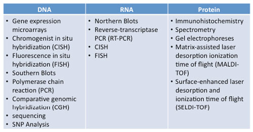
The Promise of Microarray Technology in Treating Disease"Now that you understand the concept behind array technology, picture this: a hand-held instrument that a physician could use to quickly diagnose cancer or other diseases during a routine office visit. What if that same instrument could also facilitate a personalized treatment regimen, exactly right for you? Personalized drugs. Molecular diagnostics. Integration of diagnosis and therapeutics. These are the long-term promises of microarray technology. Maybe not today or even tomorrow, but someday. For the first time, arrays offer hope for obtaining global views of biological processes-simultaneous readouts of all the body's components-by providing a systematic way to survey DNA and RNA variation." - Quote from the National Center for Biotechnology Information CISN SummaryWe have described some of the techniques used in research. It is not necessary to memorize them; they are mentioned so you have some context and if need be, you can come back to this section for details. This chart lists many of the currently used technologies arranged by tissue type utilized. Several have been described in more detail.
|
|||||||||||||||||||||||||||||||||||

1 2 |