CISN - How Cancer is Studied - Research Technology
| You Are Here: Home > Cancer Research > How Cancer is Studied > Background > Research Technology |
||||||||||||||||
Research TechnologyResearch continues to progress due to major breakthroughs in technology that allow the work to be done faster and more cheaply. Research now requires fewer cells from cheek swabs, blood samples or tissue from tumor biopsies than in the past. These samples are preserved, processed, and analyzed using a variety of techniques. Major breakthroughs in bioinformatics also allow the vast amounts of data that are collected from these types of studies to be analyzed in a way that clarifies and makes complex patterns apparent. The technologies listed below are used in all fields of cancer research: Immunohistochemistry (IHC)IHC is the process of localizing and visualizing proteins in cells of a tissue section taking advantage of the fact that antibodies bind to specific antigens. In the case of IHC, the antigen is a particular cellular protein or proteins of interest that the researcher is studying.
Gel electrophoresisThe basic principle of gel electrophoresis is that DNA, RNA, and proteins can be separated out by size from a sample mixture by means of an electric field. An agar gel containing the sample is placed in a buffer-filled box, then electrical current is applied via the power supply in the rear of the apparatus shown below. DNA and RNA naturally have a negative charge and will migrate toward a positively charged electrode. In the image shown, the negative terminal is at the far end (black wire), so positively charged DNA migrates toward the other end where the camera is located. The amount of migration will vary depending on the size (length) of the sample. Thus the pattern that shows up will be specific to the sample, analogous to a molecular fingerprint.
|
||||||||||||||||


| Section Index | |
| What We Know About Cancer | |
| ● | How Cancer is Studied |
| Drug Development | |
| New Treatments | |
| Research Advocacy | |
Southern blottingSouthern blotting is a method used for probing for the presence of a specific DNA sequence within a DNA sample. Southern blotting is less commonly used in laboratory science due to the ability of other techniques, such as PCR, to detect specific DNA sequences in samples. These blots are still used for some applications, however, such as measuring copy numbers in transgenic mice, or in the engineering of gene knockout embryonic stem cell lines. Northern blottingThe northern blot is used to study the expression patterns of a specific type of RNA molecule as relative to others in a set of different samples of RNA. Southern blot techniques are used to analyze DNA and Northern blot techniques to analyze RNA. In both cases, the nucleic acid is separated by gel electrophoresis, a process that separating molecules on the basis of their size using an electric current applied to a gel matrix. After gel electrophoresis, the material is transferred to a membrane (blot). The material is then labeled with radio-active probes that bind specifically to the pieces of the nucleic acid that are of interest. Finally, the material marked by radio-active probes is detected by exposure to an x-ray. This is shown in the picture below.
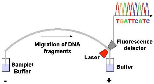
DNA sequencingThis technique was developed in 1977 by Frederick Sanger. The term DNA sequencing refers to methods for determining the order of the nucleotide bases adenine, guanine, cytosine, and thymine, in a molecule of DNA. DNA sequencing has become automated, faster and therefore cheaper. The high demand for low-cost sequencing has driven the development of high-throughput sequencing technologies that simultaneously measures multiple samples (dozens or more) in a single assay (test). The rapid speed of sequencing attained with modern DNA sequencing technology has been instrumental in allowing the Human Genome Project to sequence the human genome. In dye-terminator sequencing (image below courtesy of Wikipedia), each of the four DNA bases (T,C,A,G) is labeled with fluorescent dyes. Because each of them have different wavelengths of fluorescence and emission they produce the familiar sequence picture seen in research documents (peaks of different colors with base identified-A,T,G,C).
Owing to its greater expediency and speed, dye-terminator sequencing is now the mainstay used in automated sequencing. Knowledge of the DNA sequences of genes and other elements of the genome of organisms has become indispensable for basic research studying biological processes. Polymerase chain reaction (PCR)PCR allows a single DNA sequence to be copied millions of times (amplified), or altered in predetermined ways. There are three basic steps involved in performing a PCR. The steps are repeated 30-40 times in cycles of heating and cooling, with each step taking place at a different temperature. All of the needed components are mixed together in one tube in very tiny volumes. The reaction is carried out in an automated machine, known as a thermocycler, which is capable of rapidly increasing and decreasing the temperature. See image below.
Applications of PCR
|


1 2 |